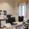
MUSAM-LAB is an experimental laboratory directed by Prof. Dr. Ing. Marco Paggi and complementing the modelling and computational facilities of the research unit MUSAM - Multi-scale Analysis of Materials.
The laboratory located in the ex-Officina Aromataria of the Convento San Francesco (XIII Century) is supported by the Starting Grant IDEAS CA2PVM from the European Research Council.
Experimental research is devoted to thermo-mechanical testing of heterogeneous materials and surfaces by investigating the complex mechanisms taking place at different scales of observation. A list of experimental failities and some illustrative applications are provided below.
Facilities
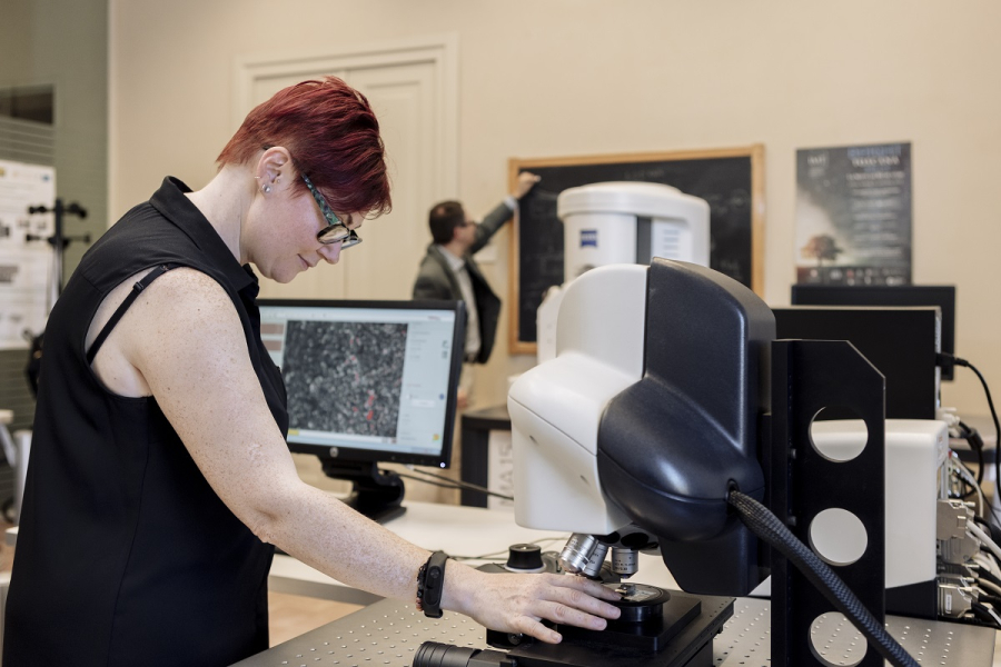
- 3D confocal-interferometric profilometer (LEICA, DCM 3D)
Non-contact optical profilometer based on confocal and interferometric methods with vertical resolution ranging from few nanometers up to several millimiters. It is able to deal with very rough but also super-smooth surfaces. Applications regard morphological characterization of surfaces and interfaces at multiple scales, analysis of coatings, wettability. Measured surface data can be treated by a research software developed in house for the statistical and fractal analysis of roughness and for the simulation of the contact response of rough surfaces (prediction of the real contact area vs. load, contact stiffness and thermal contact conductance, frictional response).
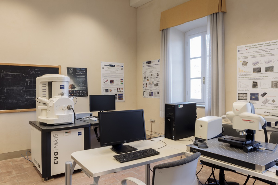
- Scanning Electron Microscope (ZEISS, EVO MA15)
SEM with dectectors for secondary and back-scattered electrons allowing a maximum resolution of 3 nm and automated pressure regulation from 10 to 400 Pa to work with metallic and also non-metallic samples without the need of metallization. The microscope can be used to visualize microstructures, defects and cracks for any kind of material (metals, ceramics, paper, polymers, etc.) at different resolutions.
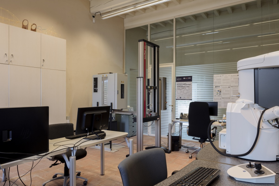
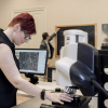
- Micromechanical testing stage (DEBEN, 5000S)
Tensile/compression stage for forces up to 5 kN specifically designed to perform mechanical tests within the SEM chamber to allow real time observation of the microstructure evolution of materials. It is also possible to test dog-bone specimens with holes or without holes, as well as compact test specimens. SEM images acquired during in-situ experimental tests can be processed according to the digital image correlation technique and compared with numerical simulations, see below.
The stage for micromechanical testing and an image of the bridging mechanism of cellulose fibers in paper tissue. - Universal testing machine with a thermostatic chamber (Zwick/Roell, Z010TH)
Universal testing machine for tensile tests, compression tests and three-point bending tests on notched beams for fracture mechanics characterization. Two load cells of 1 kN and 10 kN are available. Testing can be made inside a thermostatic chamber in the temperature range from -30°C up to 250°C.
- System for 3D displacement correlation technique (Correlated Solutions, VIC3D)
System for displacement and strain measurement using the displacement correlation technique in 3D. It can be used together with the universal testing machine as a virtual strain gauge and for 3D or 2D digital image correlation at 30 Hz. The software can also be used to process images acquired during mechanical tests inside the SEM.
- Thermocamera (FLIR, T640bx)
IR thermocamera with 640x480 pixel of resolution for thermoelastic tests in the laboratory and for in-situ inspection of hot spots in photovoltaic modules and electric circuits.
- Photocamera for electroluminesce tests (PCO, 1300 Solar)
IR photocamera specific for carrying out electroluminescence tests in the laboratory. This type of IR camera is able to detect microcracks in photovoltaic modules.
Composed of engineers, physicists and mathematicians, the unit aims at studying the deformation, fracture, fatigue, contact and the structural integrity of heterogeneous materials and structures with multi-scale and multi-physics computational and experimental methods.